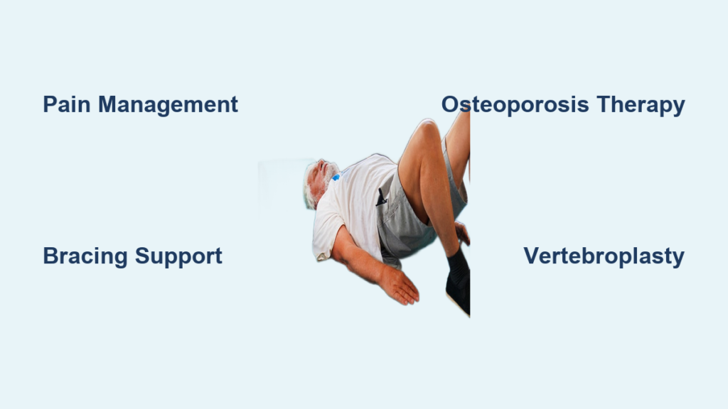
Back pain after a minor fall or even a simple sneeze might seem like a normal part of aging, but for many older adults, it can signal a compression fracture. Affecting up to half of adults over 80, vertebral compression fractures often stem from osteoporosis, where weakened bones collapse under everyday stress. Left untreated, these fractures can lead to chronic pain, loss of height, spinal deformity, and increased mortality. The good news is that timely compression fracture treatment in the elderly can restore mobility and prevent future injuries.
This guide delivers a clear, actionable roadmap for managing spinal compression fractures in older adults. From non-surgical care to minimally invasive procedures and long-term prevention, you will learn how to relieve pain, stabilize the spine, and reduce the risk of additional fractures. Whether you are a patient, caregiver, or healthcare provider, this evidence-based overview equips you with the knowledge to make confident treatment decisions.
Identifying Compression Fracture Types and Symptoms

Recognize the Three Main Fracture Patterns
Understanding the type of compression fracture guides every treatment decision. In elderly patients, fractures are classified by shape and stability:
- Wedge fracture: The most common type, accounting for more than half of all cases. The front of the vertebra collapses into a wedge shape, often from osteoporosis.
- Crush fracture: The entire vertebral body collapses uniformly on itself rather than just one side.
- Burst fracture: A severe injury where bone shatters and fragments spread in multiple directions. This type poses a significant risk to nearby nerves and requires urgent medical attention.
Fractures are also categorized as stable (fragments do not move, low nerve risk) or unstable (fragments displace, threatening nerves and soft tissues). Stable fractures typically respond to non-surgical care, while unstable ones often require surgical evaluation.
Spot the Warning Signs in Older Adults
Compression fractures in seniors may appear subtle or strike suddenly. Common symptoms include:
- Sudden back pain that worsens with standing or walking
- Gradual loss of height over time
- Kyphosis, a hunched or forward-curving posture
- Localized spinal tenderness on palpation
- Numbness, tingling, or weakness in the legs if nerves are affected
Postmenopausal women, individuals on long-term steroids, and patients with multiple myeloma face the highest risk. Even a minor fall from standing height should raise suspicion for vertebral injury.
Confirm Diagnosis With the Right Imaging
Accurate diagnosis depends on selecting the proper imaging tool:
- X-ray: First-line imaging to detect vertebral collapse, deformity, and alignment issues. Dynamic views reveal instability.
- MRI: Identifies acute fractures by showing bone marrow edema and rules out infection or tumors.
- CT scan: Provides detailed bone anatomy, especially for assessing spinal canal compromise.
- PET or bone scan: Used when malignancy is suspected.
All patients should be evaluated for underlying causes. If cancer or infection is possible, lab tests and specialist referrals are essential before finalizing a compression fracture treatment plan.
Conservative Treatment for Compression Fractures
Manage Pain Safely With Age-Appropriate Medications
Pain control is the first priority in compression fracture treatment for the elderly, but medication choices must balance relief with safety:
- Acetaminophen: First-line for mild to moderate pain.
- NSAIDs: Acceptable for short-term use in stable wedge fractures; monitor for stomach and kidney risks.
- Opioids: Reserved for severe pain at the lowest effective dose and shortest duration to avoid confusion, falls, and constipation.
- Calcitonin (Miacalcin): Intranasal spray that reduces acute fracture pain and improves mobility within days.
Always distinguish axial back pain (from bone) from radicular pain (nerve-related), since treatment protocols differ for each.
Stabilize the Spine With Bracing
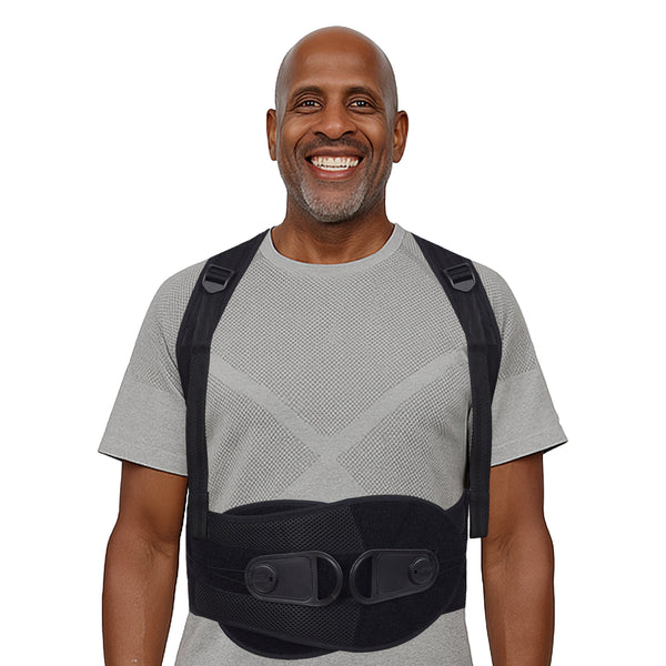
Bracing supports the spine, reduces pain, and promotes healing during the first 6 to 12 weeks.
- TLSO (Thoracic-Lumbar-Sacral Orthosis): Rigid brace most commonly prescribed for thoracic and lumbar fractures.
- Soft corsets or abdominal binders: Used later in recovery or for patients who cannot tolerate rigid bracing.
Wear the brace during activity but remove it for skin checks and hygiene. Prolonged use beyond 12 weeks can weaken core muscles, so timely weaning matters.
Begin Early Mobilization and Rehabilitation
Early movement is critical. Prolonged bed rest increases the risk of pneumonia, blood clots, pressure sores, and further bone loss.
- Ambulate within 1 to 2 days of injury, as tolerated
- Start physical therapy early, focusing on safe weight-bearing activities, core strengthening, balance training, and posture correction
- Consider inpatient rehabilitation for patients with severe pain, immobility, or neurological symptoms
Weight-bearing exercise is the cornerstone of long-term recovery and osteoporosis prevention.
Treating the Underlying Osteoporosis

Start Bone-Strengthening Therapy Immediately
Treating osteoporosis is just as important as healing the fracture itself. Without intervention, the risk of another fracture doubles.
Essential daily supplements:
– Calcium: 1,200 mg per day
– Vitamin D: 800 to 1,000 IU per day; higher doses reduce nonvertebral fractures by at least 20% in those over 65
Prescription medications to discuss with your doctor:
– Bisphosphonates (such as alendronate) to slow bone loss
– SERMs like raloxifene for postmenopausal women
– Teriparatide, a parathyroid hormone that stimulates new bone formation in severe cases
– Denosumab, an injectable that blocks bone resorption
Begin treatment as soon as medically safe, even during acute recovery.
Monitor Healing With Follow-Up Imaging
Follow-up imaging tracks healing and detects complications early.
- Repeat X-rays at 3, 6, and 12 months to assess vertebral height loss, kyphosis progression, and new fractures
- Repeat MRI if pain persists beyond 6 weeks to confirm fracture acuity or rule out other causes
Vertebral bone marrow edema typically decreases over 3 months in conservatively managed benign fractures, correlating with symptom improvement.
Minimally Invasive Procedures for Persistent Pain
Consider Vertebroplasty When Conservative Care Fails
When pain does not improve after 2 to 6 weeks of conservative treatment, vertebroplasty may be the next step.
How it works:
– Cement (polymethyl methacrylate) is injected into the fractured vertebra under X-ray guidance
– Performed under local or general anesthesia
– Most patients report pain relief within hours
Efficacy highlights:
– A Mayo Clinic review of 1,000 cases showed significant pain relief at 2 hours post-procedure
– Kim et al. reported pain improvement in 92% of 673 patients
– Best results occur in fractures with intravertebral clefts and pain lasting less than 6 months
Known risks include:
– Cement leakage (most common)
– Rib fractures during needle placement
– Adjacent-level fractures
– Rare pulmonary embolism from venous cement migration
Choose Kyphoplasty to Restore Spinal Alignment
Kyphoplasty adds a critical step before cement injection: a balloon is inflated to lift the collapsed vertebra.
Key advantages:
– Can partially restore vertebral height and reduce kyphosis
– Lower risk of cement leakage due to the contained cavity
– Faster functional recovery in select patients
Best candidates:
– Fractures within 2 to 6 weeks of injury
– Significant deformity
– Pain unresponsive to bracing and medications
Limitations to consider:
– Not suitable for severe canal compromise
– Less effective at L4/L5 due to anatomy; Han et al. found only 55.7% success at L4/L5 compared to 71.7% at L3
When Surgery Becomes Necessary
Identify Urgent Surgical Indications
Most elderly patients do well without surgery. However, immediate surgical evaluation is required when:
- Neurological deficits appear, such as leg weakness or bowel and bladder dysfunction
- Spinal instability is confirmed on imaging
- Canal compromise causes nerve compression
- Pain worsens despite conservative or minimally invasive treatment
- Deformity progresses despite bracing
These symptoms are emergencies and should never be ignored.
Understand Open Stabilization Options
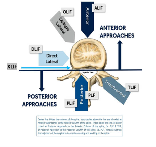
For complex fractures, open spinal fusion may be needed.
Common approaches:
– Posterior fusion: Most common technique using rods and screws to stabilize the spine
– Anterior fusion: Used for severe anterior column damage, often requiring abdominal or thoracic access
Minimally invasive spine surgery techniques are increasingly used to reduce tissue damage and speed recovery. Postoperative care typically includes bracing, inpatient rehabilitation, and a gradual return to activity.
Procedure Complications and How to Reduce Them
Watch for Cement-Related Risks
Both vertebroplasty and kyphoplasty carry risks tied to cement use:
- Cement leakage can compress nerves or enter veins, causing pulmonary embolism
- Paraparesis is a rare but serious nerve injury from misplaced cement
- Rib or pedicle fractures may occur during needle placement
Careful patient selection and skilled technique significantly reduce these risks.
Prevent Adjacent-Level Fractures After Augmentation
A major concern after cement augmentation is new fractures at nearby levels.
- Trout et al. found a 4.5 times greater risk of subsequent fracture after vertebroplasty compared to conservative care
- Adida et al. reported that two-thirds of second fractures occur at adjacent vertebral levels
- Contributing factors include severe osteoporosis, poor bone quality, and the stiffening effect of cement
Mitigation strategies:
– Aggressive osteoporosis treatment after the procedure
– Early mobilization and core strengthening
– Avoid high-impact activities during recovery
Some studies, including Zhang et al., show no increased risk, suggesting patient selection plays a key role in outcomes.
Long-Term Recovery and Prevention Strategies
Improve Prognosis With Proactive Care
Most elderly patients heal within 8 to 12 weeks with conservative treatment. Pain typically improves within 4 weeks.
Favorable outcomes depend on:
– Early diagnosis and treatment
– Prompt osteoporosis management
– Active rehabilitation
A first compression fracture signals increased frailty. Patients with concurrent vertebral and hip fractures face notably higher mortality rates, underscoring the need for comprehensive care. Pathologic fractures from cancer or infection carry a more guarded prognosis and require multidisciplinary management.
Reduce Future Fracture Risk Through Daily Habits
Prevention is the cornerstone of long-term success.
Key strategies:
– Fall prevention: Remove home hazards, install grab bars, use non-slip mats
– Balance training: Tai chi, physical therapy exercises
– Weight-bearing exercise: Walking and resistance training
– Medication adherence: Continue osteoporosis drugs as prescribed
– Regular DEXA scans: Monitor bone density over time
Patients should report new back pain immediately, avoid forward bending and heavy lifting, and ask their provider about fracture type, healing timeline, and activity limits.
Frequently Asked Questions About Compression Fracture Treatment in the Elderly
What Is the Most Common Cause of Compression Fractures in Older Adults?
Osteoporosis is the leading cause, making vertebrae too weak to withstand normal daily activities. Postmenopausal women face the highest risk due to accelerated bone loss after menopause.
How Long Does It Take for a Compression Fracture to Heal in the Elderly?
Most stable compression fractures heal within 8 to 12 weeks with conservative care. Pain typically improves significantly within the first 4 weeks, though full recovery may take longer depending on bone health and rehabilitation adherence.
When Is Surgery Needed for an Elderly Compression Fracture?
Surgery is required when neurological deficits appear, such as leg weakness, numbness, or bowel and bladder dysfunction. Spinal instability, severe canal compromise, or progressive deformity also warrant surgical evaluation.
Are Vertebroplasty and Kyphoplasty Safe for Elderly Patients?
Both procedures are generally safe when performed by experienced specialists. Studies show high success rates, with 92% of patients experiencing pain improvement after vertebroplasty. Risks include cement leakage and adjacent-level fractures, but careful patient selection reduces complications.
Can Compression Fractures Be Prevented in the Elderly?
Yes, through aggressive osteoporosis management, calcium and vitamin D supplementation, weight-bearing exercise, balance training, and home modifications to prevent falls. Regular bone density screenings help detect bone loss before fractures occur.
What Happens if a Compression Fracture Goes Untreated?
Untreated fractures can lead to chronic back pain, progressive height loss, worsening kyphosis, reduced mobility, and increased mortality. They also significantly raise the risk of additional vertebral and hip fractures.
Key Takeaways for Compression Fracture Treatment in the Elderly
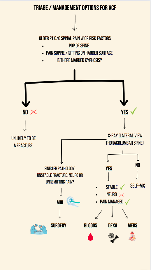
Compression fracture treatment in the elderly works best with a balanced, proactive approach. Start with safe pain management, bracing, and early mobilization, while aggressively treating the underlying osteoporosis. When conservative care fails after 2 to 6 weeks, vertebroplasty or kyphoplasty can provide rapid and lasting pain relief. Reserve surgery for neurological emergencies, instability, or progressive deformity.
The most important next step is scheduling a bone density evaluation and beginning osteoporosis therapy if you or a loved one has experienced a compression fracture. With the right combination of medical care, rehabilitation, and fall prevention, older adults can maintain mobility, independence, and a high quality of life.